Metabolomics in Metabolic Disease Research

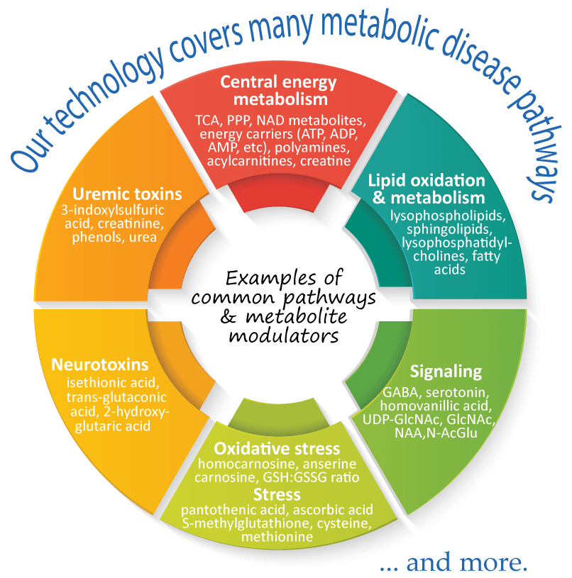
Metabolomics comprehensively characterizes small polar and lipid metabolites, yielding a snapshot of physiological processes that vary according to the pathological state of cells, tissues, and organs. Therefore, metabolomics profiling can help to reveal specific metabolic changes, for example, aberrant levels of amines, amino acids, and lipids, that are known to associate with many metabolic diseases, such as obesity, diabetes, and cardiovascular diseases. Furthermore, metabolomics has a great potential for identifying biomarkers, as well as predictive and risk markers that are associated with disease development and progression, thus, providing an effective strategy for novel therapeutic innovations.
metabolic_diseases_overview

Metabolomics brings us closer to the phenotype of an individual, providing a direct readout of metabolite alterations that occur in many metabolic diseases.


HMT’s metabolomics

HMT’s metabolome analysis employs CE-MS & LC-MS platforms. Our technologies are optimized to measure small polar metabolites that are involved in a myriad of metabolic diseases, e.g., amino acids, short-chain fatty acids, polyamines in most types of samples, including blood, tissues, and cultured cells.

Quantitation Quantitation
Over 100 polar metabolites
involved in metabolic diseases can be quantified
with single-
or multi-point calibration.
High resolution High resolution
Good separation of structural isomers, e.g. isobaric fatty acids, oxidative products.
hmt's metabolism_metabolic_diseases

Examples of samples that can be analyzed at HMT

Biofluids (plasma, serum, saliva, etc.)

  • discovery of biomarkers & diagnostic markers
  • monitor therapy-mediated changes
Urine, feces, cecal contents

  • detect inflammation-derived changes in metabolic profiles
  • screen for early diagnostic & prognostic disease biomarkers
Tissues from target organs

  • enhanced evaluation of disease progression
  • monitor therapy-mediated changes
Cultured cells

  • assessment of novel therapeutics
  • identify metabolic changes in a time-dependent manner

Recent publications

Cardiovascular Disease / Cardiac Dysfunction

1. Upregulated kynurenine pathway enzymes in aortic atherosclerotic aneurysm: macrophage kynureninase downregulates inflammation.
Nishimura et al. J Atheroscler Thromb. 2020. Epub.
2. Mitochondrial pyruvate carrier abundance mediates pathological cardiac hypertrophy.
Fernandez-Caggiano et al. Nat Metab. 2020. 2(11):1223-1231
3. Alternative oxidase-mediated respiration prevents lethal mitochondrial cardiomyopathy.
Rajendran et al. EMBO Mol Med. 2019. 11(1): e9456
4. Protein acetylation in skeletal muscle mitochondria is involved in impaired fatty acid oxidation and exercise intolerance in heart failure.
Tsuda et al. J Cachexia Sarcopenia Muscle. 2018. 9(5): 844-859
5. Titin-truncating variants affect heart function in disease cohorts and the general population.
Schafer et al. Nat Genet. 2017. 49(1): 46-53

Liver Disease / Liver Dysfunction

6. Lipid desaturation-associated endoplasmic reticulum stress regulates MYCN gene expression in hepatocellular carcinoma cells.
Qin et al. Cell Death Dis. 2020. 11(1):66
7. High throughput screening of serum γ-glutamyl dipeptides for risk assessment of nonalcoholic steatohepatitis with impaired glutathione salvage pathway.
Saoi et al. J Proteome Res. 2020. 19(7): 2689-2699
8. Effects of a DPP4 inhibitor on progression of NASH-related HCC and the p62/ Keap1/Nrf2-pentose phosphate pathway in a mouse model.
Kawaguchi et al. Liver Cancer. 2019. 8(5): 359-372
9. Canagliflozin, an SGLT2 inhibitor, attenuates the development of hepatocellular carcinoma in a mouse model of human NASH.
Shiba et al. Sci Rep. 2018. 8(1): 2362
10. Involvement of Porphyromonas gingivalis in the progression of non-alcoholic fatty liver disease.
Nakahara et al. J Gastroenterol. 2018. 53(2): 269-280

Diabetes / Obesity

11. Empagliflozin, an SGLT2 inhibitor, reduced the mortality rate after acute myocardial infarction with modification of cardiac metabolomes and antioxidants in diabetic rats.
Oshima et al. J Pharmacol Exp Ther. 2019. 368(3): 524-534
12. Identification of metabolites associated with onset of CAD in diabetic patients using CE-MS analysis: A Pilot Study.
Omori et al. J Atheroscler Thromb. 2019. 26(3): 233-245
13. Antidiabetic and cardiovascular beneficial effects of a liver-localized mitochondrial uncoupler.
Kanemoto et al. Nat Commun. 2019. 10(1): 2172
14. ACC2 deletion enhances IMCL reduction along with acetyl-CoA metabolism and improves insulin sensitivity in male mice.
Takagi et al. Endocrinology. 2018. 159(8): 3007-3019
15. CD44 variant inhibits insulin secretion in pancreatic β cells by attenuating LAT1-mediated amino acid uptake.
Kobayashi et al. Sci Rep. 2018. 8(1): 2785

Kidney Disease / Renal dysfunction

16. Hypermetabolism of glutathione, glutamate and ornithine via redox imbalance in methylglyoxal-induced peritoneal injury rats.
Hirahara et al. 2020. J Biochem. 167(2): 185-194
17. Metabolomic Changes of Human Proximal Tubular Cell Line in High Glucose Environment.
Wei et al. 2019. Sci Rep. 9(1):16617
18. Metabolomics analysis elucidates unique influences on purine / pyrimidine metabolism by xanthine oxidoreductase inhibitors in a rat model of renal ischemia-reperfusion injury.
Tani et al. 2019. Mol Med. 2019. 25(1):40
19. Metabolomic analysis of overactive bladder in male patients: Identification of potential metabolite biomarkers.
Shimura et al. Urology. 2018. 118: 158-163
20. Antioxidant modifications induced by the new metformin derivative HL156A regulate metabolic reprogramming in SAMP1/kl (-/-) mice.
Kim et al. 2018. Aging (Albany NY). 2018. 10(9):2338-2355

Other metabolic diseases

21. Progression from Monoclonal gammopathy of undetermined significance of the immunoglobulin M class (IgM-MGUS) to Waldenstrom Macroglobulinemia is associated with an alteration in lipid metabolism.
Jalali et al. Redox Biol. 2021. 41:101927
22. Novel metabolic disturbances in marginal vitamin B6-deficient rat heart.
Kumrungsee et al. J Nutr Biochem. 2019. 65:26-34./td>
23. Loss of the E3 ubiquitin ligase MKRN1 represses diet-induced metabolic syndrome through AMPK activation.
Lee et al. Nat Commun. 2018. 9(1): 3404
24. A skeletal muscle model of infantile-onset Pompe disease with patient-specific iPS cells.
Yoshida et al. Sci Rep. 2017. 7(1): 13473
25. The mammalian malonyl-CoA synthetase ACSF3 is required for mitochondrial protein malonylation and metabolic efficiency.
Bowman et al. Cell Chem Biol. 2017. 24(6): 673-684

Resources

For more detailed information, visit our Resources page where you can download the following:

Download 

Home